Best experienced on desktop

You can browse scans on your phone. Large studies, 3D views, and detailed measurements work best on a desktop; mobile performance and controls may be limited.

Choose a tool, then drag on the image. Use the strip below the image to move through slices.

Read the whole scan. Your agent can, too.

Any WebMCP-enabled agent, such as Codex, can bring its own intelligence to the scan and use every tool you can.

Handles
CT, MR, PET, X-ray
Opens
A folder, a ZIP, or a disc
Needs
No install, no account, no upload
Agents
WebMCP-enabled, such as Codex
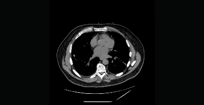
Chest CT

Lung and soft-tissue stacks, coronal and sagittal reconstructions, MIP, and scouts.

Noncontrast and contrast axial images, with coronal and sagittal reconstructions.

KU Leuven cadaveric specimen 7. Distal forearm, wrist and hand with axial, coronal and sagittal derived series; supports bone reconstruction.

Cadaveric knee: sagittal, coronal and axial imaging plus 3D DESS. Five selected sequences with original pixel detail.

3D T1 and T2, postcontrast T1, and FLAIR. This source examination does not include DWI/ADC.

Colorized PET and fused PET/CT over the CT’s shorter field of view. Explore 419 PET slices, linked planes, activity values, and 3D MIP.

Two chest projection images. Source metadata does not label individual view positions.

Three shoulder projections: axial and internal/external-rotation AP views.

CC BY 4.0Source & attribution for Chest CT (opens in a new tab)

Nothing is uploaded. Your browser opens the files itself and keeps them here until you remove them. Interpretation happens in the agent you ask, not on this device.

WebMCP lets agents such as Codex review the scan.

Codex gets first-class access to the same tools you do, so it works through the images itself instead of guessing from a screenshot.

How to try it

  1. Open the Codex desktop app
  2. Open this page within the in-app browser
  3. Ask for what you want in the chat:
    • Review and interpret this scan. Annotate and provide key images where needed.
    • Show me the gallbladder on axial and coronal views.
    • Measure the displacement of the fracture.

All studies

Sources & attribution

What the agent can do

Codex reaches this viewer through 26 WebMCP tools. Those tools let it:

Look through the study

  • List the studies on this device, and open one
  • Browse the series in a study and step to any image
  • Change the plane, layout, zoom and window
  • Reformat to coronal, sagittal, oblique or 3D
  • Put two studies or series side by side

Read the actual data

  • Read the value at a point in real units, Hounsfield for CT
  • Sample voxels across a region
  • Measure distances and angles in millimeters from the scan geometry
  • List every measurement already on the study

Leave a record

  • Save key images, and reopen the exact view any of them came from
  • Pin observations to a point in the study
  • Draft a report with findings, impression and limitations
  • Cite the key images a report is based on

Stay accountable

  • Undo what it just did
  • Download the original DICOM files it worked from

Remove imported study?

Remove “” and its saved observations from this browser? Your source files are unaffected.